Sample analysis of dike rocks.
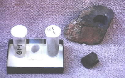
Specimen G1-1 is shown above. It is an oriented sample of the contact between the basaltic dike (the black right side of the specimen) and the syenite bedrock (the coarse-grained, lighter left side of the specimen). The basalt is fine-grained because the intrusion was chilled when the melt was injected into the cooler, older syenite. The basalt has been drilled to produce a cylinder for the paleomagnetic portion of the study. Shown also are two vials of crushed sample for the chemical analysis, and a thin section of the rock between the vials. The thin section is used for the analysis under a petrographic microscope.
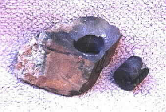
The view above shows sample G1-1 after the core has been drilled for the paleomagnetic study. This paleomagnetic study was initiated by the late S. K. Runcorn who was then a visiting scholar at Penn State from the University of Newcastle-Upon-Tyne. This study was the first work involving paleomagnetism by a graduate student at Penn State. Because at that time Penn State did not have the equipment to support paleomagnetic research, I drilled the cores, prepared the samples, and ran the initial analysis at the University of Pittsburgh, and then continued the analysis at the plaeomagnetic laboratories at Princeton University. Paleomagnetism is the determination of the direction of the north magnetic pole when the rock was formed. Because, relative to any continent, the magnetic poles move throughout time, the magnetic pole position can be used to infer the age of the rock.
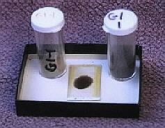
The thin section between the two vials is used for a petrographic analysis. I made the above thin section, as well as all others which were used in the study. However, the dike rocks were highly altered by late-stage hydrothermal solutions, so the petrology revealed under the microscope was compromised. Consequently, I resorted to bulk chemical analyses to determine the compositions of the different types of dikes.
The vials above contain crushed sample for the chemical analyses, with one vial containing coarse crushing fraction and the other contains the fine fraction. For a chemical analysis, the specimen must be crushed sufficiently fine that the entire portion will pass through a 200-mesh nylon sieve. The finely crushed samples are then mixed with a flux (lithium metaborate), fused in a furnace, rapidly quenched into a glass bead, dissolved in a strong acid, spiked with a cobalt internal standard, and analyzed using flame photometry and emission spectroscopy.